Overview
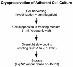
Cryopreservation of adherent cell cultures requires detaching cells from the culture surface, typically by trypsinization or another appropriate cell dissociation method. The detached cells are collected by centrifugation, gently resuspended in an appropriate cryopreservation medium at the desired viable cell density, and aliquoted into sterile cryogenic vials. The cryovials are then frozen using a controlled-rate cooling method (approximately −1°C/min), typically by placing them in a controlled-rate freezing device housed in a −80°C freezer overnight. Alternatively, other validated controlled-rate freezing methods may be used. Following controlled-rate freezing, the cryovials are transferred to liquid nitrogen or a −150°C freezer for long-term storage.
Note: This protocol provides a general procedure for cryopreserving adherent mammalian cell cultures. Cryopreservation requirements may vary among cell lines. Therefore, always consult the manufacturer’s recommendations or supplier’s instructions for cell line-specific protocols to ensure optimal post-thaw recovery and viability.
 Requirements
Reagents
♦ Growth medium (e.g., DMEM supplemented with 10% FBS)
♦ PBS (Ca2+-free and Mg2+-free)
♦ Trypsin-EDTA solution (or another suitable cell dissociation reagent)
♦ Cryopreservation (freezing) medium (e.g., 90% complete growth medium + 10% DMSO, or 80% complete growth medium + 10% FBS + 10% DMSO, depending on the cell line)
Equipment and disposables
♦ Sterile cryogenic vials
♦ Sterile conical tubes (15 mL or 50 mL) (e.g., SARSTEDT, 62.554.502 or 62.547.254)
♦ Controlled‑Rate Freezing Container/−80°C freezer or programmable Controlled‑Rate Freezers
♦ Hemocytometer and trypan blue solution (or an automated cell counter)
♦ -80°C freezer
♦ Liquid nitrogen storage system or −150°C freezer
♦ Benchtop centrifuge with 45° fixed-angle or swinging-bucket rotor (e.g., Eppendorf™ 5804 Series)
♦ Laminar Flow Hood
♦ Micropipettees and sterile pipette tips
♦ Serological pipettes and pipette controller
♦ Inverted Phase‑Contrast Microscope
♦ Appropriate personal protective equipment (laboratory coat, gloves, and cryogenic face/eye protection)
Starting materials
Healthy adherent cultures in the late logarithmic growth phase, approximately 80–90% confluent, with low passage number whenever possible.
Tip: Replace the culture medium one day before cryopreservation to remove cellular debris and ensure that the culture is healthy and actively proliferating.
Protocol
Prior to starting
1. Carefully inspect the cell culture under an inverted phase-contrast microscope to ensure that the cells are healthy and free of contamination.
2. Confirm that the culture is mycoplasma-free before cryopreservation.
3. Label each cryogenic vial with the cell line name, passage number, date, and operator initials (recommended).
Related articles:
- Mycoplasma Contamination in Cell Culture
- Detection of Mycoplasma Contamination
- Sources of Mycoplasma Contamination
- Effects of Mycoplasma on Cultured Cells
Step 1: Harvest cells from the culture dish by trypsinization
- Aspirate the culture medium and wash the monolayer with calcium- and magnesium-free PBS.
- Add sufficient trypsin-EDTA solution to cover the cell layer (typically 1–2 mL for a T25 flask) and incubate at 37°C for approximately 1–2 minutes.
- Observe cell detachment microscopically. If necessary, continue incubation until the cells detach completely.
- Gently tap the flask to release any remaining adherent cells.
- Add serum-containing complete growth medium (approximately 4 mL for a T25 flask) to neutralize trypsin.
- Pipette gently across the culture surface to recover all cells.
- Transfer the cell suspension to a sterile centrifuge tube.
Note:
Serum in the complete growth medium has trypsin inactivating activity.
Tips:
1. At this stage, cells can be collected by centrifugation, and the resulting cell pellet can be resuspended in an adequate amount of growth medium. However, this is optional. If you suspect significant cell death during trypsinization or if the cell suspension is too diluted, centrifugation and resuspension must be performed.
2. Cell suspensions from cultures of the same cell line and passage number may be pooled.
Cautions:
1. Avoid prolonged exposure to trypsin, as excessive enzymatic treatment may decrease cell viability and reduce post-thaw recovery.
2. Ensure that all cells are completely recovered from the culture vessel. Incomplete cell harvesting can reduce the total cell yield and may adversely affect the number of cryovials prepared.
Step 2: Determine viable cell density in cell suspension (optional)
- Mix 20 μL of cell suspension with 20 μL trypan blue solution.
- Load the mixture onto a hemocytometer.
- Count viable (unstained) and non-viable (blue-stained) cells.
- Calculate the viable cell concentration (cells/mL) and cell viability (%).
Tips
1. Automated cell counters (e.g., Countess® Automated Cell Counter or Moxi Flow Kit) can be used as an alternative.
2. If the suspension is overly dilute, centrifuge the cells and resuspend the pellet in a smaller volume.
3. Cell counting is particularly important for primary cultures or other limited cell populations.
4. Experienced users may estimate the expected number of cryogenic vials from routine cultures; however, cell counting provides greater accuracy.
Cautions
1. Ensure that the cells are properly resuspended before counting the viable cells.
2. Cell viability should preferably exceed 90% before cryopreservation. Cultures with viability below 80–85% generally exhibit poorer post-thaw recovery.
3. Avoid cryopreserving cultures with poor viability whenever possible. Instead, expand the culture under optimal conditions before freezing.
Step 3: Resuspend cells in ice-cold freezing medium at the recommended viable cell density of (1 x 10⁶ – 5 x 10⁶ cells/ml)
- Harvest the cells from the cell suspension by centrifugation at 4°C for 5 to 10 minutes at 250-300 × g (1000 – 1500 rpm for the Eppendorf™ 5804 Series benchtop Centrifuge).
- Carefully aspirate the supernatant as much as possible without disturbing the cell pellet.
- Flick the tube with your finger several times to dislodge the pellet.
- Add an appropriate volume of ice-cold freezing medium to achieve the desired viable cell density (between 1 x 10⁶ – 5 x 10⁶ cells/ml). Resuspend the cells thoroughly using gentle pipetting.
Tip
Cell density in the freezing medium can vary by cell line. In most cases, a higher cell density is beneficial for cell recovery.
Cautions
1. Dimethyl Sulfoxide (DMSO) is cytotoxic, so exposure time should be minimised. Use a chilled freezing medium to reduce its harmful effects, and keep the cell suspension at 4 °C throughout handling.
2. Quickly resuspend the pellet in the freezing medium immediately after aspirating the supernatant.
3. Centrifugation speed and duration must be carefully optimised to obtain a soft, loose pellet that retains the maximum number of cells. If the centrifugation force is too low, a significant portion of the cells may remain suspended and be lost during supernatant removal. Conversely, excessive speed or prolonged centrifugation produces an overly compact pellet that is difficult to resuspend. Tight pellets often require vigorous pipetting to break apart, and this mechanical stress can damage cell membranes, reduce viability, and compromise experimental outcomes. For this reason, centrifugation conditions should always be selected to achieve a balance: firm enough to collect the cells efficiently, yet gentle enough to preserve pellet softness and ensure easy, low‑shear resuspension.
Step 4: Aliquot the cell suspension into cryogenic vials.
- Place labeled cryovials on ice.
- Dispense 1 mL of cell suspension into each cryovial.
- Immediately tighten the vial caps securely.
Caution
Gently mix the cell suspension periodically during aliquoting to maintain a homogeneous cell concentration among all cryovials.
Step 5: Subject cryovials to slow cooling (1 – 3°C/min) overnight in -80°C freezer
Place all cryogenic vials in a controlled-rate freezing container (e.g., CoolCell® Cell Freezing Containers from Biocision) and store immediately in a -80°C freezer overnight.
Tips
1. The most efficient method for freezing cryogenic vials is to use a controlled‑rate freezer, such as the CryoMed™ Controlled‑Rate Freezers from Thermo Fisher Scientific. These instruments precisely regulate the cooling profile, typically at −1 °C per minute, ensuring optimal dehydration and ice‑crystal formation dynamics that preserve cell viability. However, for most serum‑grown cell lines, specialised controlled‑rate equipment is not strictly necessary. Commercially available passive cooling devices—such as the CoolCell® Cell Freezing Containers from Biocision or the Mr. Frosty™ Freezing Container from Thermo Fisher Scientific—provide a reliable and reproducible cooling rate suitable for routine cryopreservation. These devices use isopropanol‑based or insulated designs to achieve an approximate −1 °C per minute cooling rate when placed in a −80 °C freezer, making them practical and cost‑effective alternatives for standard laboratory workflows.
2. Homemade insulated containers (small size thermocol boxes filled with cotton or tissue paper) may be used when commercial devices are unavailable; however, cooling rates may be less reproducible.
Step 6: Store cryogenic vials in liquid nitrogen
- Transfer all frozen cryogenic vials to a liquid nitrogen container the next day. Alternatively, they can also be stored in a -150°C freezer.
- Cryogenic vials can be stored in either the gas phase or the liquid phase of liquid nitrogen.
Tip
A frozen vial can be revived after two weeks to confirm that the frozen stocks are viable and free from contamination.
Caution
1. Biosafety Level 2 (BSL-2) cell lines should be stored in the gas phase of liquid nitrogen.
2. It is important to maintain accurate records of the locations of frozen cell lines.
3. Wear protective equipment when handling liquid nitrogen. Be aware that cryogenic vials may explode if stored in the liquid phase of liquid nitrogen.
Further reading:
- Freshney, R. I., 2010. Basic Cell Culture Protocols / Culture of Animal Cells: A Manual of Basic Technique and Specialized Applications (6th ed.). Wiley-Blackwell.
- Stacey, G. N., & Masters, J. R., 2008. Cryopreservation and banking of cell lines. Nature Protocols, 439, 215–232. PMID: 19180080, DOI: 10.1038/nprot.2008.190, Nature
Reliability Index *****
Note: We welcome your feedback. If you notice any errors, inconsistencies, or have suggestions for improvement, please share your comments in the box below. Your feedback helps us continuously improve the quality, accuracy, and usefulness of our content.
Highest reliability: *****
Lowest reliability: *****
Disclaimer: While we strive to provide accurate and up-to-date information, we cannot guarantee its absolute accuracy or completeness. The information contained on this website is for general informational purposes only and should not be considered as professional advice. We disclaim any liability for any loss or damage resulting from the use of the information provided herein. Always consult qualified professionals for specific guidance. Read more

2 thoughts on “Protocol – Cryopreservation of Adherent Cell Culture”